
Fundus Autofluorescence And Optical Coherence Tomography Of A Macular Cherry Red Spot In A Case Report Of Sialidosis Bmc Ophthalmology Full Text

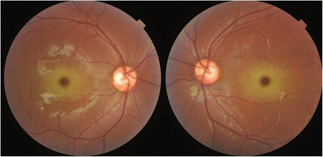
Left Eye Showing Cherry Red Spot With Retinal Pallor Typical Of Central Download Scientific Diagram
3
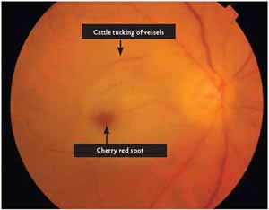
Hollenhorst Plaque With Branch Retinal Artery Occlusion Causing A Cherry Red Spot
Cherry On The Top
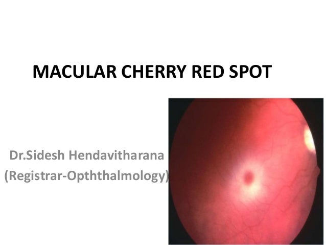
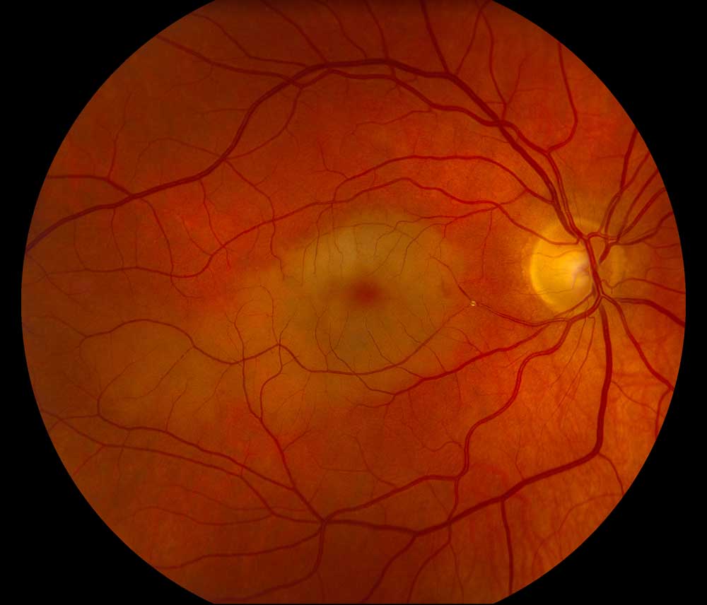
Macular Cherry Red Spot

Cherry Red Spot Telltale Sign Of Neurometabolic Disorders Global Genes

Scielo Brasil Cherry Red Spot In A Patient With Tay Sachs Disease Case Report Cherry Red Spot In A Patient With Tay Sachs Disease Case Report
Differential Diagnosis Of Cherry Red Spot Medical Zone

View Image
1
Tags:
Archive


